
Image
Snapshot quiz (May 2021 [2])
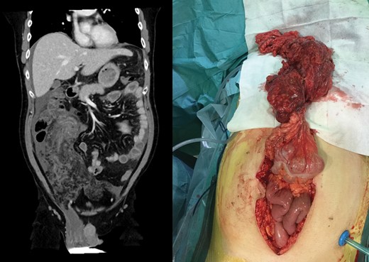
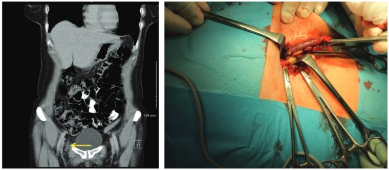
A 35-year-old man presented with a 4-day history of abdominal pain and fever. Abdominal examination showed a distended abdomen with signs of localized peritonitis in the right iliac fossa. Admission blood tests included leucocytosis of 24.5 × 10⁹/l and a C-reactive protein level of 450 mg/l. What is shown on the CT image and accompanying intraoperative photograph?
View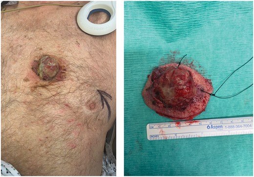
Image
Snapshot quiz (May 2021 [1])
An 84-year-old man presented to the acute surgical intake with a rapidly growing painless lesion on the chest wall. He had an implantable pacemaker and defibrillator inserted a year previously. What is the diagnosis and management?
View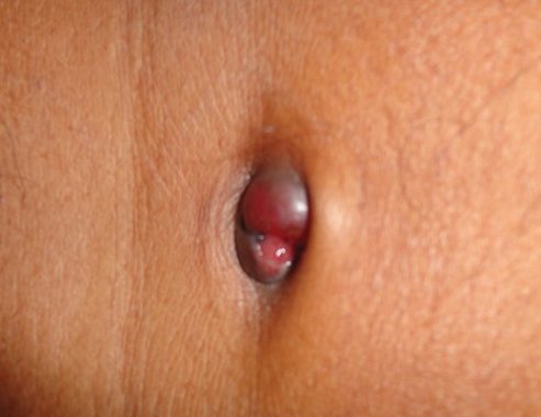
Image
Snapshot quiz (April 2021 [1])
A 42-year-old woman presented with a 3-month history of dyspepsia, weight loss, and vague upper abdominal pain. On examination a palpable umbilical nodule was found. What is this clinical sign called?
View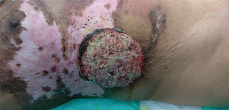
Image
Snapshot quiz (January 2021)
A 45-year-old woman with a history of an old burn injury to the chest and abdominal wall presented with a progressive ulcerated lesion on her right flank. What is the diagnosis?
View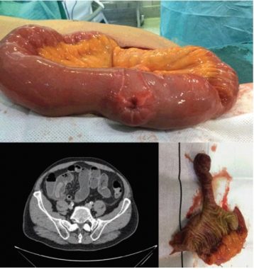
Image
Snapshot quiz 20/12
A 65-year-old man with no previous surgical history presented with acute abdominal pain, vomiting, anaemia and a raised C-reactive protein level. CT was done and the patient was taken to the operating theatre. What is the diagnosis?
View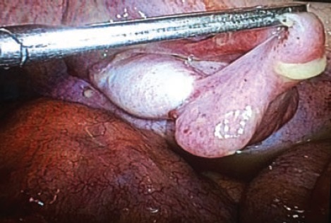
Image
Snapshot quiz 20/11
During diagnostic laparoscopy in a 50-year-old patient, the following intra-abdominal structure was found near the internal inguinal ring. What is the diagnosis?
View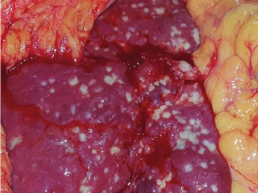
Image
Snapshot quiz 20/7
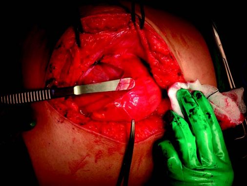
A 70-year-old man presented with right upper quadrant abdominal pain and fever. Levels of serum inflammatory markers and obstructive hepatic enzymes were raised. A colectomy had previously been performed for cancer. The photograph shows the liver at laparotomy. What is the diagnosis and what is the usual clinical presentation?
View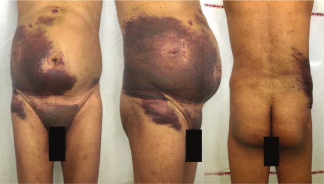
Image
Snapshot quiz 20/6
What is the condition in this 68‐year‐old man following a laparoscopic cholecystectomy?
View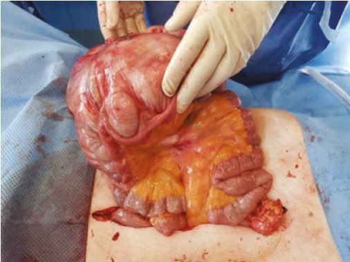
Image
Snapshot quiz 20/4
An elderly man presented with a rapidly enlarging right iliac fossa mass. CT demonstrated a 20-cm well circumscribed lobulated mass, intimately related to the caecum and terminal ileum. Biopsy was not diagnostic. During surgery it was seen that the mass appeared to arise from the mesentery. What is the likely diagnosis?
View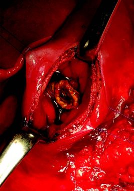
Image
Snapshot quiz 19/12
This patient had undergone distal pancreatic resection for pancreatic cancer, including resection of the coeliac trunk and portal vein. The portal vein was reconstructed with an artificial prosthesis. What has happened?
View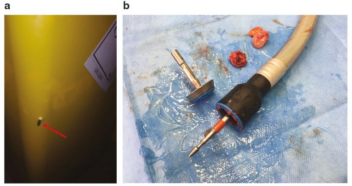
Image
Snapshots in surgery: A cautionary tale
A member of the theatre team had a superficial injury to her leg from the spike of a circular stapler (CDH series, Ethicon Endosurgery, Cincinnati, Ohio, USA) that has protruded through a sharps bin (a). Here, the surgeon did not retract the spike before disposal in an overfilled bin. All team members must take care when disposing of circular staplers, beginning with the surgeon retracting the spike immediately after checking the tissue ‘doughnuts’ (b).
View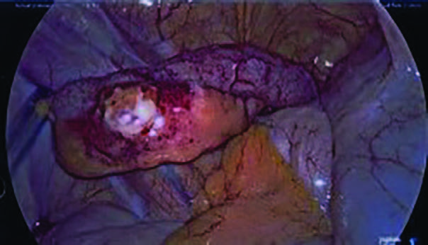
Image
Snapshot quiz 19/9
This patient presented with appendicitis. What is the diagnosis and how should it be managed?
View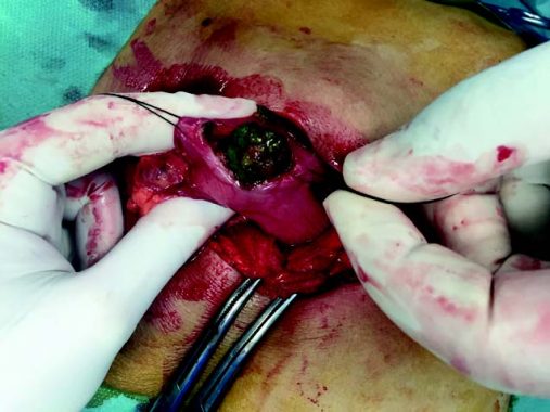
Image
Snapshot quiz 19/7
This 50-year-old woman, with no previous abdominal surgery, presented with symptoms of intestinal obstruction for 3 days. Since the age of 16 years she had experienced recurrent abdominal colic. What is the cause of intestinal obstruction here?
View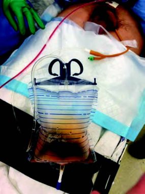
Image
Snapshot quiz 19/6
A 35-year-old man underwent laparoscopic appendicectomy for perforated acute appendicitis. A drain was placed via the suprapubic trocar at the end of surgery. After operation, the urinary catheter bag was found to be filled with air. What is the diagnosis?
View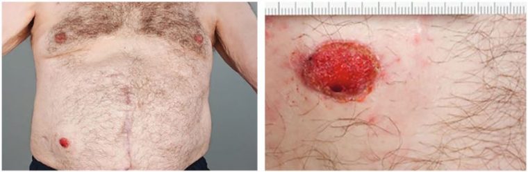
Image
Snapshot quiz 19/5
What is this abdominal lesion in a 78-year-old man having surveillance for colorectal malignancy?
View
Image
Snapshot quiz 19/1
What is the cause of these liver capsule adhesions found during laparoscopy?
ViewThe Association of Surgeons in Training was originally established in 1976 and is the second largest surgical specialty association in the UK with over 2,700 members, from all ten surgical specialties.
Image
Snapshot quiz 18/18
What type of hernia is this, shown on the CT scan (left) and operative illustration (right)?
View